Dr. Jane Ruby joined Stew Peters with revelation regarding the ‘magnetism’ videos circulating in social media of people sticking metal objects or magnets to themselves. The CDC published on their site “receiving the ‘vaccine’ will not make you magnetic, including at the site of ‘vaccination’ which is usually your arm.” Dr. Ruby says, “They’re lying”, and provided evidence in support of her claim, as well as a disturbing warning about the ingredients in the jab, directly from the source.
Brittany Galvin can stick metal object on her face
Brittany Galvin joins ‘The Stew Peters Show’ LIVE from her Florida hospital bed, where she is undergoing treatment for a serious diagnosis that, according to her neurologist, was caused by the Moderna injection. www.StewPeters.tv
CDC denies the vaccine contains metal particles that can make people magnetic
The CDC website published a page titled Myths and Facts about COVID-19 Vaccines where the following appears

Are Magnetic Nano particle real and are they applied in medicine?
An article published in the Journal of Nanobiotechnology ( part of Springer Nature) gives a good overview of a variety of magnetic nano particles, their use and application in the biomedical field where they can be used both in the body and outside the body. There seems to be main 4 groups of application discussed in research literature : Magnetic separation of biological entities like purification of blood, Magnetic nanocarriers contributed to drug delivery, Radio frequency-controlled magnetic nanoparticles provided a new approach for cancer treatment and Magnetic nanosensors for resonance imaging (MRI) applications. However I am sure the unpublished innovations are lightyears a head of all that is published.
According to Magnetic Nanoparticles: From Design and Synthesis to Real World Applications published on 29 August 2017 Synthesis of magnetic nanoparticles used for medical and biomedical applications are widely available commercially
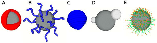
Figure 2. Structures of magnetic particles and their coating schemes. (A) Core-shell magnetic particle; (B) End-grafted polymer coated magnetic nanoparticle; (C) Magnetic particle fully encapsulated in polymer coating; (D) Heterodimer magnetic particle; (E) Hydrophobic magnetic particle encapsulated within lipid monolayer (upper part) and hydrophilic magnetic particle within lipid bilayer (modified from [41] with permission).”
Can nano particles be magnetic without containing metal nano particles and have they been used in mRNA therapeutics?
Magnetic Nanoparticles: From Design and Synthesis to Real World Applications section 4.4. Cells and Biomolecules reads:
“Another strategy to support DNA interaction with magnetic particles is to induce positively charged moieties and promote electrostatic interaction with a negatively charged phosphate nucleic acids backbone. It has been demonstrated that surfaces modified with amino groups provide a positive charge in acidic conditions due to amino groups’ protonation and additionally provide electrostatic interaction without additional agents like chaotropes. It was recognized that the density of amino groups positively influences ability to adsorb DNA. Such an increase can be obtained using particles with a rough surface or amine-functionalized mesoporous silica magnetic particles [172,173]. Commonly, the sol-gel method is used for magnetic particle silanization. In subsequent steps, APTES containing a –NH2 group is induced. Alternatively, polyethyleneimine (PEI) as a molecule with a high density of amine moieties can be used for particle modification [174]. Polycharged magnetic particles modified by a surface-coating either with silica, polyvinylpyrrolidone or tripolyphosphate are easy to make in the laboratory and have demonstrated interactions with DNA for magnetic isolation applications [38]. Sequence specific isolation of nucleic acids can be performed using magnetic particles decorated with oligonucleotide probes which recognize target nucleic acid according to its sequence [175,176]. Adams et al., compared abilities of commercially available magnetic particles for isolation of RNA—silica coated, particles decorated with a polythymine oligonucleotide probe (oligo (dT)) and particles functionalized with an oligonucleotide complementary to target sequence [177]. Total mRNA was selectively extracted from the real sample spiked with target mRNA using oligo (dT). Silica coated particles isolated total RNA and particles with specific probe extracted mRNA highly enriched with the target sequence but exhibited significantly slower binding kinetics.
The previously discussed methods are used for peptides/protein enrichment through nonspecific hydrophobic interactions with carbon materials, silica, n-alkyl or mesoporous silica [178,179,180,181]. However, methods for specific peptide/protein isolation are rather developed. It can be achieved using antibody or MIPs immobilization on magnetic material [182,183]. Conversely, a hydrophilic surface of such particles is desired to minimize nonspecific adsorption. The imprinting of biomolecules is problematic in comparison with small molecules since they can denature during the fabrication process. Recombinant proteins with a polyhistidine tag can be efficiently purified using their affinity to metal ions like Ni2+. Although immobilized metal affinity chromatography (IMAC) and metal oxide affinity chromatography (MOAC) are standardly used for their purification, several magnetic materials exploiting this powerful affinity were reported [184,185]. Similar mechanism is used also for separation of phosphorylated protein/peptides [186,187].
Therapeutic delivery of nucleic acids into a patient’s cells as an alternative to drugs is of importance nowadays. However, low cell membrane translocation efficiency, short in vivo half-time and poor cell-specific targeting, complicates its broader application. In this field, delivery of microRNA (miRNA) is a highly investigated area. miRNAs are short noncoding RNA molecules which interact with target mRNAs to down-regulate or inhibit translation. Enhanced apoptosis of cancer cells induced by the combined effect of hyperthermia and miRNA delivery by magnetic particles was described by Yin et al. [241]. They targeted heat shock proteins Hsp70 and Hsp90 which protect cellular proteins from degradation and hinder of hyperthermia-induced apoptosis [242]. Mesoporous silica magnetic particles were used as a carrier for small interfering RNA (siRNA) for the down-regulation of vascular endothelial growth factor gene [243]. Further, they were capped by PEI-PEG cover to increase their biocompatibility and by fusogenic KALA Amphipathic Peptide (WEAKLAKALAKALAKHLAKALAKALKACEA) to increase their cell internalization.”
Magnetic Nanoparticles: From Design and Synthesis to Real World Applications
More on the magnetic properties of silica can be found in the following paper MAGNETIC PROPERTIES OF SILICA WITH MESOPORES STRUCTURED AS MCM-48
Seeing it first hand on someone I know
I don’t know what magnetic nano particles are used in the mRNA vaccines since not all the ingredients are disclosed. But what I do know is that the magnet do stick to peoples bodies and I had witnessed it first hand. Here is my little experiment: I was in a zoom conversation with a young person who got the Pfizer vaccine. Without too much introduction or fun fair I asked her if she was aware of the magnet challenge, she had no idea what I was talking about. I asked her if she has a magnet in her room, she said yes. Go get it I said. When she got back with the magnet I asked to place it in the vaccination site on her arm. And it did stick. She was shocked! She then tried to place it on the other arm and it stuck as well. I was surprised by that. At this point I was not aware that was possible.
What are the risks of using Magnetic Nano Particles internally in humans?
An article published on the Biochemical functionality of magnetic particles as nanosensors: how far away are we to implement them into clinical practice? brings together information from 114 studies related to this topic here are some tidbits :
- SPIONs = superparamagnetic iron oxide nanoparticles
- MNPs = magnetic nanoparticle
“SPIONs are generally composed of γ-Fe2O3 or Fe3O4. In comparison to other metal and metal-oxide nanoparticles, SPIONs have the advantage of being compatible in a biological environment [34]. Also, they undergo biodegradation [34]. This has made SPIONs prominent candidates for in vivo applications. Biodegradation of SPIONs is dependent on coating and coating material as well as on size.Coating influences biodegradation due to partial hindered access to the metal-oxide core [35]. Concerning biodegradation, very small particles (< 20 nm) will be quickly eliminated in the body by the kidneys, whereas on the other hand large nanoparticles (> 200 nm) will be filtered in the liver and spleen [36]. These are important aspects when aiming at an in vivo application of the nanoparticles.
The main difference between carbon-coated MNPs and SPIONs,from a pure materials point of view, is the higher saturation magnetization of the former,which leads to a much faster separation of carbon-coated MNPs when applying a magnetic field. Additionally, SPIONs are superparamagnetic while carbon-coated MNPs are ferromagnetic [37]. This means that carbon-coated MNPs have a tendency to aggregate due to their permanent magnetization, which may impose a challenge when using bare carbon-coated MNPs for in vivo applications.However, aggregation may be hindered by surface modification of the carbon-coated MNPs to produce stable dispersions [38]. Furthermore, magnetic properties of nanoparticles can also be influenced by other factors than the material choice such as size of the particles, crystallinity, shape and composition [39]. This should be considered when tailoring nanoparticles towards specific properties and applications.
Physicochemical characteristics of the synthesized magnetic nanosensors determine biocompatibility. As a consequence, interactions with the biological milieu such as the blood have to be considered carefully. Blood is a complex liquid consisting of different molecular and cellular entities. Therefore, it is important to ensure that magnetic nanosensors do not interfere with blood in any other way than intended, nor should they induce any unwanted reactions such as inflammation. In general, a suitable coating with a polymer is sufficient to achieve biocompatibility before the sensing functionality is implemented (Fig. 1). A widely applied approach is dextran coating of nanosensors used as contrast agents for MRI [40,41,42]. Another possibility to coat MNPs with a polymer is the use of atomic transfer radical polymerization (ATRP), a technique, which allows the production of a variety of polymers [43]. This approach is utilized to polymerize functionalized methacrylate onto carbon-coated cobalt nanoparticles in order to achieve stable dispersions of MNPs [38]. These functionalized nanoparticles have an azide moiety, which may be further modified by “click”-reaction to include substrates that may be used for biomedical applications [38]. An additional biocompatible polymer is polyglycerol (PG). Polyglycerol has a chain structure similar to polyethylene glycol (PEG). The advantage of PG is its optimal hydrophilicity, stability and resistance to non-specific adsorption of proteins [44]. A simple one-step synthetic approach for PG is anionic ring opening polymerization, which results in a hyperbranched polymer. Biocompatibility of such PG was tested. Results revealed similar or even better behaviour of PG compared to PEG [45, 46]. Recently, hyperbranched PG was polymerized onto MNPs (Fe2O3). As a consequence, MNPs have become resistant to nonspecific adsorption of proteins [47]. Due to the simple synthesis process, the biocompatibility as well as the possibility for further functionalization, PG coating is a valuable alternative approach for the preparation of magnetic nanosensors for biomedical applications.
Various studies exist assessing possible toxic effects of MNPs in a living organism. Surface characteristics determine distribution within the body, whereas size, dose and entry point of nanoparticles are important as well. In general, inflammation may be triggered through stimulation of effector cells, producing proinflammatory mediators, whereas the proinflammatory effect seems to be surface-dependant [94]. When MNPs remain in the tissue over time, chronic inflammation may be another consequence leading to fibrosis of the affected organ [95, 96]. Finally, MNPs may evoke damage, which triggers the development of cancer [97]. This is of particular concern as long-term studies are still missing.
Nanoparticles, once present in the body, may target various systems. There are effects of nanoparticles found on the circulatory system, where nanoparticles indirectly influence for example blood pressure [98]. Important to note when looking at the circulatory system is the fact that nanoparticles are engineered to influence the coagulation system of the blood [99]. At the same time, MNPs, designed for any other indication, may evoke an unwanted pro- or anticoagulant effect in the blood [100].
The reproductive system is another target of nanoparticles with possible detrimental effects.Upon in vivo applications, nanoparticles may accumulate in reproductive organs [102, 103], where they have direct effects on germ cells with reduced cell count or activity in both, female and male germ cells [104, 105]. Furthermore, nanoparticles are able to alter or damage DNA in cells, which would be especially problematic in germ cells [106]. In a recent in vitro study, uptake of coated SPIONs in granulosa cells was tested. It was found that depending on the coating, no or only low uptake and toxicity of SPIONs was observed [107].”
Interestingly many adverse effects of this technology mirror the adverse effects of the mRNA vaccines reported by many world wide.


